Human Anatomy and Physiology
- 28.4K likes on YouTube
- 1.5M views
Created by S R Pharmacy
What you'll learn
- Human Anatomy and Physiology • Cell structure and definition
- Human Anatomy and Physiology • Introduction to anatomy and physiology
- Levels of organisation: cells, tissues, organs, systems
- Types of connective tissue
- Muscular tissue types
- Skeletal system overview
- Physiology • Composition of blood
- Physiology • Antigens on red blood cells
Explore related topics
Course content
01Structure of cell || HAP chapter 2 || Components of cell || Human anatomy and physiology chapter 2Preview32:22
This chapter from Human Anatomy and Physiology (Chapter 2, Structure of the Cell) defines the cell as the basic structural and functional unit of living organisms and the building block of the body. Cells form tissues, tissues form organs, organs form systems, and systems make up the whole body. The cell carries out many functions such as energy production, protein and lipid synthesis, and keeping the organism alive.
- Human Anatomy and Physiology • Cell structure and definition
- Human Anatomy and Physiology • Cell membrane and cell wall
- Human Anatomy and Physiology • Nucleus and genetic material
- Human Anatomy and Physiology • Mitochondria and energy production
- Human Anatomy and Physiology • Endoplasmic reticulum, Golgi complex and lysosomes
- Human Anatomy and Physiology • Prokaryotic vs eukaryotic cells
02Human Anatomy and Physiology chapter 1 || Chapter first Human Anatomy and Physiology || HAPPreview24:55
This chapter introduces Human Anatomy and Physiology (HAP). Anatomy is the branch of science that studies the structure, shape, position, location and relationships of body parts, such as bones, muscles, nerves and organs. Physiology is the branch that studies the functions of body parts and how they work, including metabolism, digestion, and the pumping action of the heart. In short, anatomy describes what a structure is and where it lies, while physiology explains what it does.
- Human Anatomy and Physiology • Introduction to anatomy and physiology
- Human Anatomy and Physiology • Scope of the subject
- Human Anatomy and Physiology • Directional terms
- Human Anatomy and Physiology • Body planes
- Human Anatomy and Physiology • Body cavities
03HAP Chapter 3 || Tissues of the human body || Epithelial | Connective || Muscular || Nervous tissuesPreview16:27
This chapter gives an overview of the tissues of the human body, following the chain of organisation from cells to tissues, organs, and systems. Cells are the basic building blocks of the body. A group of cells with similar structure and function forms a tissue, several tissues combine to form an organ (the liver, for example, contains epithelial, connective and nervous tissue), and several organs working together form a system, such as the digestive system. Many systems together make up the whole body.
- Levels of organisation: cells, tissues, organs, systems
- Epithelial tissue types
- Connective tissue functions
- Muscular tissue
- Nervous tissue
04Connective Tissue || HAP chapter 3 part 2 || Types of connective tissuePreview25:58
This chapter, part 2 of the Human Anatomy and Physiology (HAP) Chapter 3 series, covers connective tissue. Connective tissue supports and connects other body tissues and organs, holds them in place, and stops them from moving or shifting. Its main types are fibrous connective tissue, bone tissue, blood, and cartilage. Fibrous connective tissue has three main forms: loose (areolar) fibrous tissue, which lies under the skin and around organs, blood vessels, nerves and mucous membranes, and is a gel-like semi-fluid matrix containing fibres and cells such as fibroblasts, adipocytes, macrophages and mast cells; dense irregular fibrous tissue, found in the dermis and organ capsules; and dense regular fibrous tissue, with parallel fibres, found in tendons and ligaments. Tendons join muscle to bone, while ligaments join bone to bone. Reticular tissue, made of reticular fibres forming a net-like framework, is found in the spleen, lymph nodes and bone marrow, and helps make blood cells and filter microorganisms.
- Types of connective tissue
- Loose and dense fibrous connective tissue
- Reticular tissue and its functions
- Bone tissue: compact and spongy bone
- Cartilage types and locations
- Blood as a connective tissue
05Muscular Tissue || Nervous Tissue || types of muscular tissue || HAP Chapter 3 part 3Preview31:04
This chapter (Human Anatomy and Physiology, Chapter 3 part 3) covers two tissue types: muscular tissue and nervous tissue. Muscular tissue is responsible for contraction and relaxation, movement of the skeletal system, locomotion of the body, and generating force and heat. It has three types. Skeletal muscle is attached to bones by tendons, is made of long cylindrical multinucleated fibres containing actin and myosin, is striated, is voluntary, and is controlled by the somatic nervous system. Smooth muscle is non-striated, has single nuclei, is involuntary, is found in the walls of internal organs and hollow structures such as blood vessels, the gut, urinary tract and reproductive organs, and is controlled by the autonomic nervous system. Its functions include peristalsis, vasoconstriction and vasodilation, and uterine contraction. Cardiac muscle is found in the myocardium of the heart, is striated with a single nucleus per cell, has many mitochondria because it works continuously, shows continuous and rhythmic contraction, is involuntary, and pumps blood.
- Muscular tissue types
- Skeletal muscle
- Smooth muscle
- Cardiac muscle
- Nervous tissue and neurons
- Glial cells and neuron types
06Human Anatomy and physiology chapter 4 || Osseous system || Skeleton System || disorders of JointsPreview46:32
The skeletal system, also called the osseous or bony system, is the body's framework of bones, joints and associated tissues. Its basic structural unit is the osteon, and it is studied in two parts. The axial skeleton is the central axis: the skull (29 bones), the vertebral column (26 bones) and the rib cage (25 bones, including the sternum). The appendicular skeleton is the limbs and their girdles, with 126 bones. A newborn has about 270 bones, which fuse during growth to about 206 in adults. The count of 206 is the standard figure, though some very small bones are not counted. The axial skeleton has 80 bones and the appendicular skeleton 126, giving 206 in total. The skull has 8 cranial bones and 14 facial bones, plus 6 ear ossicles (3 per ear). The hyoid bone is also counted in the skull. The vertebral column has 7 cervical, 12 thoracic, 5 lumbar, 5 fused sacral and 1 coccygeal region. The skeleton protects organs, supports the body, enables movement, stores minerals such as calcium and phosphorus, and helps form blood cells.
- Skeletal system overview
- Axial and appendicular skeleton
- Bone count and regions
- Classification of joints
- Joint movement types
- Joint disorders
07Haemopoietic system || HAP chapter 5 || Process of Hemopoiesis || Mechanism of Blood ClottingPreview33:45
This chapter introduces the haemopoietic (haematopoietic) system, the organs and tissues such as bone marrow, spleen, thymus, lymph nodes and liver that help form blood cells. Blood is a liquid connective tissue that transports oxygen, carbon dioxide, nutrients, wastes, and helps regulate temperature and clotting. Blood is made of plasma (about 55%), a liquid containing about 92% water plus proteins such as albumin and globulin, electrolytes, glucose, amino acids, vitamins, hormones and antibodies; and blood cells (about 45%). The three main cell types are red blood cells (erythrocytes, rich in haemoglobin, about 4.5 to 5.5 million per microlitre, lifespan about 120 days, no nucleus), white blood cells (leukocytes, about 4500 to 11000 per microlitre, granulocytes such as neutrophils, eosinophils and basophils, and agranulocytes such as lymphocytes and monocytes, which fight infection and perform phagocytosis), and platelets (thrombocytes, 1.5 to 4.5 lakh per microlitre, lifespan about 10 days, essential for clotting). Cancers of red cells, white cells and platelets are polycythaemia, leukaemia and thrombocythaemia respectively, all originating in bone marrow.
- Physiology • Composition of blood
- Physiology • Blood cells and their functions
- Physiology • Haemopoiesis
- Physiology • Mechanism of blood clotting
- Physiology • Disorders of blood
08Blood Grouping || Importance of blood GroupingPreview28:00
This chapter (Human Anatomy and Physiology, Chapter 5 Part 2) explains blood grouping and why it matters. Blood groups are decided by antigens, which are proteins on the surface (outer membrane) of red blood cells (RBCs). Based on which antigens are present, RBCs fall into four groups: A (antigen A only), B (antigen B only), AB (both A and B) and O (no antigen). A person has only one of these patterns in their RBCs, not a mix of A and B on separate cells. The Rh (Rhesus) factor is a separate antigen that adds the positive or negative label, so groups are written as A positive, A negative, and so on. Blood grouping is done by mixing the sample with reagents and seeing whether the blood clumps.
- Physiology • Antigens on red blood cells
- Physiology • ABO blood group system
- Physiology • Rh (Rhesus) factor
- Physiology • Plasma antibodies and compatibility
- Physiology • Universal donor and recipient
- Physiology • Importance of blood grouping in transfusion
09HAP chapter 6 || Lymphatic system || Lymph || Structure and functions of spleen and lymph nodePreview26:10
This chapter covers the lymphatic system from the HAP (Human Anatomy and Physiology) syllabus, focusing on lymph, lymph nodes and the spleen. Lymph is a watery, clear fluid that flows through lymphatic vessels. It forms from blood plasma by diffusion: as blood moves through capillaries, water, proteins, lymphocytes and some other components pass out into the surrounding tissue space. Lymph is about 94% water, with proteins, fats, carbohydrates, urea, creatinine, lymphocytes and minerals such as sodium, calcium, potassium and chlorine. Its functions include supplying nutrition and oxygen to areas that blood does not reach directly, maintaining the amount of tissue fluid, carrying fat from the intestine to the blood, helping in destruction of foreign particles, transporting metabolic wastes from tissues to blood, and producing lymphocytes. The lymphatic system is a network of lymphatic vessels, lymph nodes and organs such as the spleen, thymus, bone marrow and tonsils.
- Lymph formation and composition
- Functions of lymph
- Lymphatic system organisation
- Structure and functions of lymph nodes
- Structure and functions of spleen
10#human_heart #hap_chapter_7 full lecture k liye playlist dekhiye #srpharmacyPreview00:58
This chapter introduces the structure of the human heart, shown as a beating heart in the lecture. The heart has four chambers: two upper chambers called atria and two lower, larger chambers called ventricles. The right atrium receives deoxygenated blood from the body through two large veins: the superior vena cava, which brings blood from the upper body, and the inferior vena cava, which brings blood from the lower body. The left atrium is on the other side and receives oxygenated blood from the lungs through the pulmonary veins. The lecture notes that the two upper chambers can be seen separately from the outside, while the two lower chambers cannot be clearly separated when viewed from the front.
- Human Physiology • Heart chambers
- Human Physiology • Atria and ventricles
- Human Physiology • Venae cavae
- Human Physiology • Pulmonary veins and oxygenated blood
11Hap chapter 7 || Cardiovascular System || anatomy and physiology of heart || Blood vesselsPreview37:41
This chapter introduces the cardiovascular system, made up of the heart and blood vessels, and covers the anatomy and physiology of the heart. The heart has four chambers: two upper atria (right and left) that receive blood, and two lower ventricles (right and left) that pump blood out. Four valves keep blood flowing in one direction: the tricuspid valve (right atrium to right ventricle), the pulmonary valve (right ventricle to pulmonary artery), the mitral or bicuspid valve (left atrium to left ventricle), and the aortic valve (left ventricle to aorta). The heart wall has three layers: the outer epicardium (pericardium), the middle myocardium (muscle), and the inner endocardium. Blood vessels are arteries (carry blood away from the heart), veins (carry blood toward the heart), and capillaries (the thinnest vessels, where exchange happens). Arteries are not defined only by oxygen content: pulmonary artery carries deoxygenated blood, and pulmonary veins carry oxygenated blood.
- Heart chambers and valves
- Heart wall layers
- Blood vessels: arteries, veins, capillaries
- Cardiac conduction system
- Cardiac cycle and blood pressure
- Blood circulation: pulmonary, systemic, coronary
12Basics of ECG || Blood pressure and its regulationPreview42:24
This chapter introduces the basics of the electrocardiogram (ECG) and blood pressure. An ECG is a test of heart health that records the heart's electrical activity using an electrocardiograph; it is taken with electrodes placed on the four limbs (limb leads) and six points on the chest (chest leads). Electrical impulses start at the SA node, spread through the atria and then the ventricles, and each spread causes contraction (depolarisation) followed by relaxation (repolarisation). On the ECG these events appear as the P wave (atrial depolarisation), the QRS complex (ventricular depolarisation and contraction) and the T wave (ventricular repolarisation and relaxation). Changes in these waves help identify conditions such as angina, where ST-segment depression appears, a serious heart attack, where ST-segment elevation appears, and atrial fibrillation, where the baseline is irregular and the P wave disappears.
- Physiology • ECG basics and waves
- Physiology • Conduction system of the heart
- Physiology • Cardiac cycle and circulation
- Physiology • Blood pressure and its measurement
- Physiology • Cardiac conditions on ECG
13Respiratory System || HAP chapter 8Preview41:00
The respiratory system is responsible for the exchange of gases, mainly oxygen and carbon dioxide, between the body and the external environment. The process of exchange is called respiration. External respiration is the exchange of gases between the atmosphere and the blood, which happens in the lungs. Internal (tissue or cellular) respiration is the exchange between the blood and the body's cells. Air travels through the nose or mouth, then the pharynx, larynx (voice box), trachea, bronchi, bronchioles and finally the alveoli. Gas exchange happens across the thin walls of the alveoli by diffusion, which moves gases from higher to lower concentration. Oxygen moves into the blood and carbon dioxide moves out. Red blood cells (RBCs) carry the gases in the blood, with haemoglobin binding oxygen.
- Respiratory system and gas exchange
- Respiratory organs and their functions
- Mechanism of breathing
- Lung volumes and capacities
14Digestive System || human anatomy and physiology chapter 9 || HAP chapter 9 || part 1Preview1:06:48
The digestive system is a set of organs and tissues that work together to convert ingested food into simple forms that the blood can absorb. Digestion has two kinds: mechanical (chewing, churning, grinding, without enzymes) and chemical (enzymes and juices break food into smaller units). The alimentary canal runs from the mouth through the pharynx, oesophagus, stomach, small intestine (duodenum, jejunum, ileum), large intestine (caecum, appendix, colon parts, rectum) and anus. Accessory organs such as salivary glands, liver, gall bladder and pancreas help digestion without being part of the canal.
- Digestive system organs and alimentary canal
- Digestion: mechanical vs chemical
- Saliva and salivary digestion of starch
- Stomach and gastric juice
- Small intestine, pancreatic juice and bile
- Liver, gall bladder and pancreas functions
15Physiology of absorption || types of absorption || hap chapter 9 part 2 || important questionsPreview15:51
This chapter (Physiology of Absorption, Human Physiology Chapter 9 Part 2) explains how digested food is moved from the gastrointestinal tract into the blood. Absorption is defined as the movement of food, or any substance, from the GI tract into the blood circulation or blood vessels. Once absorbed, nutrients travel through the blood to supply the body's needs such as energy, glucose, fats, vitamins and minerals. Digestion must come first, because only small, simple molecules can pass through the gut wall.
- Physiology • Absorption definition
- Physiology • Passive diffusion and filtration
- Physiology • Endocytosis
- Physiology • Facilitated diffusion and active transport
- Physiology • Digestive system and exam structure
16HAP Important Questions 2024Preview07:21
This chapter covers the core topics of Human Anatomy and Physiology (HAP) through typical exam questions. It starts with the structure and classification of the body: types of joints and their locations, the parts of a human cell, and the classification of body tissues, especially epithelial and connective tissue. It then moves to the cardiovascular and respiratory systems. Key ideas include the anatomy and function of the heart, the cardiac cycle with its systole and diastole phases, blood pressure and how the body regulates it, the blood clotting mechanism, and the mechanics of breathing, where the diaphragm and chest muscles change lung volume. Lung volumes such as tidal volume, vital capacity and residual volume are also defined.
- Anatomy • Joints and tissues
- Physiology • Cardiovascular system
- Physiology • Respiratory system
- Physiology • Digestive system and saliva
- Physiology • Nervous system and reflexes
- Physiology • Endocrine system
17HAP important questions and answers 2024 || part 1Preview50:03
This chapter is a revision session on Human Anatomy and Physiology (HAP) covering four long-answer topics. First, joints are classified by movement: immovable (fixed, such as skull sutures), slightly movable (such as joints between vertebrae), and freely movable (ball and socket, hinge, pivot, gliding, saddle, and double hinge joints), with examples and common disorders such as arthritis, gout, bursitis and dislocation. Second, the structure of a human cell is described: the plasma membrane controls what enters and leaves, the nucleus controls cell activities and holds DNA, the endoplasmic reticulum (rough one makes proteins with ribosomes, smooth one is for other work), lysosomes digest material, the cytoplasm is a fluid that holds organelles, and mitochondria make ATP. Prokaryotic cells lack a well-defined nucleus, while eukaryotic cells have one and membrane-bound organelles. Third, the four tissue types (epithelial, connective, muscular and nervous) are described, with epithelial tissues classified as simple squamous, stratified squamous, simple columnar and simple cuboidal, and connective tissues (areolar, adipose, reticular and dense) are explained. Fourth, the heart and circulation are covered: its four chambers, valves, the flow of blood through pulmonary and systemic circulation, the conduction system (SA and AV nodes), the cardiac cycle, systolic and diastolic pressure, normal blood pressure values and how it is measured. The chapter ends with the mechanism of breathing, where inspiration and expiration change lung pressure relative to the atmosphere.
- Human Anatomy • Joints and their classification
- Human Biology • Structure of a cell
- Human Biology • Tissues of the human body
- Human Physiology • Heart and cardiac cycle
- Human Physiology • Blood pressure
- Human Physiology • Mechanism of breathing
18HAP important Questions and answers 2024 || part 2 || most important questions of HAPPreview1:01:00
This chapter (HAP important questions, part 2) covers several human anatomy and physiology topics. It starts with the digestive system: the parts of the gastrointestinal tract (mouth, pharynx, oesophagus, stomach, duodenum, jejunum, ileum, caecum, appendix, colon, rectum, anus) and accessory organs (liver, pancreas, gallbladder). Digestion is explained as mechanical (teeth, in the mouth only) and chemical (enzymes and acids). Salivary amylase starts carbohydrate digestion in the mouth, pepsin acts on proteins in the stomach, and bile, pancreatic juice and intestinal enzymes (amylase, trypsin, lipase) complete digestion and absorption in the small intestine. Saliva's composition and functions (moistening, lubrication, taste, starting digestion) are also covered.
- Human Anatomy • Digestive system and digestion
- Physiology • Menstrual cycle
- Physiology • Muscle contraction
- Physiology • Autonomic nervous system responses
- Physiology • Renin-angiotensin-aldosterone system
- Physiology • Endocrine glands and pituitary hormones
- Physiology • Spermatogenesis
19HAP most important questions and answers (Short) || Hap important short questionsPreview42:25
This chapter is a short-answer revision session on Human Anatomy and Physiology (HAP). It opens with the scope of anatomy and physiology, which helps in understanding the structure and function of the human body, and explains why doctors, physiotherapists and drug developers need this knowledge. The blood section covers its composition (plasma plus cells such as RBCs, WBCs and platelets) and its functions, mainly transport of nutrients, oxygen, carbon dioxide and wastes, along with temperature regulation, clotting, storage and defence. Blood clotting is explained as a cascade: platelets release thromboplastin, which with calcium converts prothrombin to thrombin, thrombin converts fibrinogen to fibrin threads, and these trap blood cells to seal the wound. The spleen (RBC formation in fetal life, blood storage, lymphocyte formation and filtering of foreign particles) and the cardiac cycle are also covered.
- Anatomy & Physiology • Scope and importance
- Anatomy & Physiology • Blood composition and blood clotting
- Anatomy & Physiology • Respiratory volumes and control of breathing
- Anatomy & Physiology • Reflex arc, hearing and skin
- Anatomy & Physiology • Urine formation and micturition
- Anatomy & Physiology • Endocrine glands and lymphatic system
20HAP chapter 10 || Skeletal muscle || Physiology of muscle contractionPreview21:05
This chapter covers skeletal muscle, a type of tissue attached to bones by tendons. Skeletal muscle is made of long, cylindrical, multinucleated muscle fibres. Each fibre contains myofibrils, which are built from repeating contractile units called sarcomeres. The fibre is covered by a membrane called the sarcolemma. Thick filaments are made of myosin, with myosin heads that project outward. Thin filaments are made of actin, along with tropomyosin and troponin.
- Human Physiology • Skeletal muscle structure
- Human Physiology • Sarcomere and myofilaments
- Human Physiology • Mechanism of muscle contraction
- Human Physiology • Role of calcium, ATP and troponin
- Human Physiology • Disorders of skeletal muscle
21Nervous System || HAP chapter 11 || Part 1Preview25:59
This part of Chapter 11 (Nervous System, Biology, Class 11/12 level) introduces the nervous system as the body's control and coordination system. It works through a network of neurons (nerve cells) that carry signals, called impulses, between the brain, spinal cord and body parts. Vital functions like heartbeat and breathing run automatically, while voluntary actions like moving a hand are under conscious control. The nervous system is first divided into the central nervous system (brain and spinal cord) and the peripheral nervous system (12 pairs of cranial nerves and 31 pairs of spinal nerves that connect the CNS to organs and tissues).
- Nervous system organisation
- Central and peripheral nervous system
- Autonomic nervous system
- Parts of the brain
- Functions of the cerebrum and cerebellum
- Functions of the medulla, mid brain and hypothalamus
22Anatomy and physiology of sympathetic and parasympathetic nervous System || hap chapter 11 part 2Preview30:30
This chapter (Human Anatomy and Physiology, chapter 11, part 2) covers the spinal cord, reflexes, cranial nerves, and the autonomic nervous system. The spinal cord runs inside the vertebral column and gives rise to 31 pairs of spinal nerves, one from each segment: 8 cervical, 12 thoracic, 5 lumbar, 5 sacral and 1 coccygeal. Reflex actions are automatic, rapid responses controlled by the spinal cord without involving the brain. A reflex arc is the circuit made of a receptor, a sensory neuron, an integration centre in the spinal cord, a motor neuron, and an effector that produces the response, as when a hand is pulled away from a hot object.
- Spinal cord and spinal nerves
- Reflex action and reflex arc
- Cranial nerves
- Autonomic nervous system overview
- Sympathetic nervous system
- Parasympathetic nervous system
23Sense Organs || HAP chapter 12 || eye || skin || ear || nose || tonguePreview27:10
This chapter covers four sense organs (eye, skin, ear and tongue) plus the nose, focusing on the structure and physiology of each. The eye is a ball-shaped organ. Light enters through the transparent cornea, passes through the pupil (an opening formed by the iris), and is focused by the lens. The ciliary muscles change the lens shape to focus on near or far objects. Aqueous humor fills the space in front of the lens, and vitreous humor fills the large space behind it. The choroid carries blood vessels that supply nutrition and oxygen, and the sclera is the tough white outer coat. The retina contains photoreceptors: rods detect light and movement, and cones detect colour. The retina converts light into electrical impulses that travel along the optic nerve to the brain, where they are interpreted as an image.
- Structure and function of the eye
- Physiology of hearing
- Structure and functions of the skin
- Taste and tongue physiology
- Olfaction and the nose
- Sense organs overview
24Urinary System || HAP chapter 13Preview31:59
This chapter from the HAP (Human Anatomy and Physiology) course covers the urinary system, also called the renal system. Its main job is to filter waste products from the blood and remove them from the body as urine. It also regulates electrolyte balance (sodium, chloride, magnesium, potassium), body fluid and blood volume, and blood pressure. The system consists of a pair of kidneys, a pair of ureters, the urinary bladder and the urethra. Blood reaches the kidneys through the renal artery and leaves through the renal vein. Each kidney has an outer cortex, an inner medulla and a covering renal capsule. The nephron is the basic structural and functional unit of the kidney. Its parts are the glomerulus, Bowman's capsule, the proximal convoluted tubule (PCT), the loop of Henle and the distal convoluted tubule (DCT), which drain into the collecting duct. The bladder is a temporary store for about half a litre of urine. The urethra carries urine out of the bladder; in males it is also the common passage for semen.
- Urinary system anatomy
- Nephron structure
- Urine formation
- Micturition (urination reflex)
- Renin-angiotensin system
- Kidney function tests
25HAP Chapter 14 || Endocrine System || Human Anatomy and Physiology Chapter 14Preview39:32
The endocrine system is made of endocrine glands, which release chemical substances (hormones, or secretions) directly into the blood, unlike exocrine glands, which release their secretions through ducts to a target site such as the skin or the lining of an organ. Examples of exocrine glands include salivary glands, sweat glands, and sebaceous glands. The chapter covers the main endocrine glands: the pituitary, thyroid, parathyroid, adrenal, pancreas, and gonads, plus a brief mention of the pineal gland.
- Endocrine glands vs exocrine glands
- Anterior and posterior pituitary hormones
- Thyroid and parathyroid hormones and calcium regulation
- Adrenal gland hormones
- Pancreatic hormones insulin and glucagon
- Gonadal hormones (testosterone, estrogen, progesterone)
26Reproductive System || HAP Chapter 15Preview1:04:53
This chapter from the HAP (Human Anatomy and Physiology) course, titled Reproductive System, introduces how the male and female reproductive systems work together to make reproduction possible. It begins with male anatomy: the testes (which produce sperm and sit in the scrotum, kept slightly cooler than body temperature), the epididymis (a coiled tube where sperm mature and are stored), the vas deferens (a duct carrying mature sperm toward the urethra), the seminal vesicles and prostate gland (which together make most of the semen fluid, rich in fructose and enzymes that nourish and protect sperm), the bulbourethral (Cowper's) glands (which release a lubricating fluid into the urethra), and the penis with the urethra running through it. Spermatogenesis is the formation of sperm from germ cells in the seminiferous tubules, helped by Sertoli cells and regulated by FSH, LH and testosterone.
- Male reproductive system anatomy
- Spermatogenesis
- Female reproductive system anatomy
- Menstrual cycle phases
- Oogenesis and ovulation
- Pregnancy and parturition
27Human Anatomy and Physiology Whole syllabus Quick Revision today at 2 : 00 PM || Be ready at timePreview2:14:50
This revision lecture covers a wide sweep of Human Anatomy and Physiology. Anatomy describes body structure (what and where), while physiology describes how body parts function. The cell is the basic structural and functional unit of living organisms; cells group into tissues, tissues into organs, and organs into systems. Key cell organelles include the nucleus, mitochondria (the powerhouse that produces ATP), endoplasmic reticulum (rough ER makes proteins), Golgi apparatus (packages and stores proteins), and lysosomes (digestive enzymes). The four main tissue types are epithelial (covering and protection), connective (binding and support, including blood, bone and fibres), muscular (movement, including skeletal, smooth and cardiac muscle), and nervous (control and coordination via neurons).
- Cell structure and organelles
- Tissues of the human body
- Skeletal system and joints
- Blood and blood groups
- Cardiovascular and lymphatic systems
- Respiration, digestion and nervous system
Description
For D Pharma first year
About the creator

- YouTube channel
- 184K subscribers
- 3 courses on Studyio
The videos in this course are made by S R Pharmacy and play from YouTube, credited to them. Studyio adds the chapters, notes, quizzes and a certificate around them.
More from S R Pharmacy

More courses like this
Optimal Protocols for Studying & Learning

Business Studies Class 12

Manzil 2026: Complete CHEMISTRY Playlist
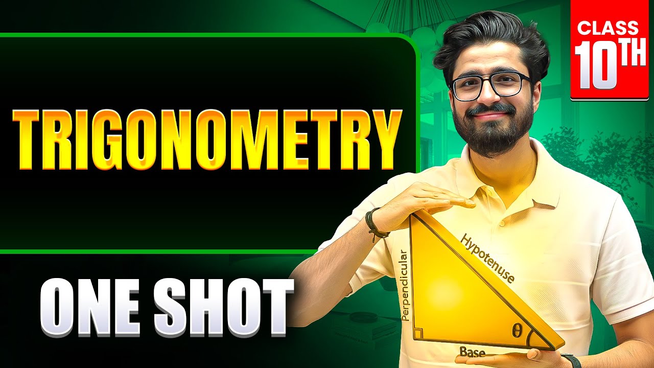
Trigonometry ONE SHOT 🔥 | Class 10 Maths Chapter 8 | Ritik Mishra
UNIT AND DIMENSION Unit and Dimensions FULL CHAPTER | Class 11th Chapter 1 | Arjuna JEE
